Gut bacteria influence the progression of multiple myeloma

Gut bacteria influence the progression of multiple myeloma

By interacting with the immune system, some types of gut bacteria can influence the progression of multiple myeloma, a tumor that affects the bone marrow causing pain, anemia and bone fragility.

The discovery, limited so far to the animal model of the disease, is published today in Nature Communications by the team of Matteo Bellone, head of Cellular immunology Unit at IRCCS Ospedale San Raffaele. The study, supported by AIRC – The Italian Association for Cancer Research – is among the first to trace a direct link between intestinal microbiota and a tumor located in a different organ, thus demonstrating the capability of these bacteria to interact with the whole organism. Moreover, researchers identified a biological marker that could predict the aggressiveness of multiple myeloma in asymptomatic patients and suggest the efficacy of some anti-inflammatory drugs, already approved for other conditions, in slowing down the disease progression.

Multiple myeloma is a severe tumor affecting plasma cells, the immune cells responsible for antibody production. In patients with multiple myeloma, these cells accumulate inside the bone marrow, interfering with its normal blood regeneration activity and weakening the bones. The disease is preceded by a painless and asymptomatic phase, in which some plasma cells have already acquired tumor characteristics and release a specific protein that can be detected in both the blood and the urine of patients, even in the absence of other pathological manifestations.

To understand the process underlying the disease progression from its asymptomatic phase to the symptomatic one, researchers focused on the animal model of the disease and on the role played by commensal bacteria. They discovered that a specific type of bacteria, also present in humans and called Prevotella heparinolytica, promotes the multiplication of inflammatory lymphocytes Th17 and that these lymphocytes are directly involved in tumor progression inside the bone marrow.

According to the study, Th17 cells migrate from the gut to the bone marrow, where they foster plasma cells proliferation, facilitating the transition from the asymptomatic phase of multiple myeloma to the actual disease through the release of a cytokine – an inflammatory molecule – called IL-17. «Given the key role IL-17 plays in multiple myeloma progression, this molecule could become a predictive tool: the amount of IL-17 in the bone marrow of asymptomatic patients could be the first biomarker able to identify patients at high risk of developing multiple myeloma», explains Arianna Brevi, first author of the research together with Arianna Calcinotto.

To test their hypothesis on the role of Th17 lymphocytes and IL-17 cytokine, researchers performed two experiments: at first, they blocked IL-17 and other inflammatory molecules

involved in tumor progression using anti-inflammatory drugs already approved for other conditions; secondly, they modified mice commensal microbiota, through the administration of antibiotics and the transplantation of bacteria species of known anti-inflammatory activity. In both cases, they managed to slow down the onset of multiple myeloma.

“The evidence collected suggests a way to identify patients at greater risk of developing multiple myeloma and to act in advance, thus containing the disease in its asymptomatic stage or, at least, slowing down its more severe manifestation”, says Matteo Bellone. “Although the experimental results obtained so far have to be further confirmed in clinical settings, they provide new hopes for patients and clinicians.”

Story Source: http://research.hsr.it/en/news/gut-bacteria-influence-the-progression-of-multiple-myeloma.html

More information: Microbiota-driven interleukin-17-prodcuing cells and eosinophils synergize to accelerate multiple myeloma progression, Nature Communications doi.org/10.1038/s41467-018-07305-8


Continue Reading

Related Articles

Antibiotics Affect the Efficacy of Immunotherapy

Antibiotics Affect the Efficacy of Immunotherapy

A study published in the journal Science by a research team from Gustave Roussy, INSERM, INRA, AP-HP, IHU Médiaterranée Infections* and Paris-Sud University shows that prescribed antibiotics impair the efficacy of immunotherapy in cancer patients.

It is important to consider that more than 20% of patients living with cancer receive antibiotics. The authors explored patients’ gut microbiota composition by metagenomic analysis and demonstrated that the bacterium Akkermansia muciniphila was associated with a better clinical response to anti-PD-1 antibody immunotherapy. Moreover, oral administration of this bacterium to mice with an unfavorable microbiota restored the anti- tumor activity of the immunotherapy.

Immunotherapy represents a real revolution in cancer therapies and has been shown to be superior to standard chemotherapy in advanced melanoma, lung, renal and bladder cancer. Although a large proportion of patients still do not benefit from this treatment, “Our research partially explains why some patients do not respond. Taking antibiotics has a deleterious impact on survival in patients receiving immunotherapy. Furthermore, the composition of the intestinal microbiota is a new predictive factor for success,” summarized Dr. Bertrand Routy, hematologist and member of the team of Professor Laurence Zitvogel, director of the “Immunology of tumors and immunotherapy” laboratory (Inserm/Paris-Sud University/Gustave Roussy).

In a cohort of 249 patients treated with anti-PD-1/PD-L1 based immunotherapy for advanced lung, kidney or bladder cancer, 28% received antibiotics for minor infections (dental, urinary or lung infections) but their general health status was not different from patients not receiving antibiotics. The study’s findings revealed that taking antibiotics two months before and up to one month after the first treatment had a negative effect on progression-free survival and/or overall survival for these three types of cancer.

The precise composition of the gut microbiota was established by metagenomics both before and during immunotherapy in 153 patients with advanced lung or kidney cancer. The identification of all the bacterial genes present in the gut microbiota was performed by INRA (MetaGenoPolis, Dr. Emmanuelle Le Chatelier). A favorable microbiota composition, rich in Akkermansia muciniphila, was found in patients with the best clinical response to immunotherapy and in those whose disease had not progressed for at least 3 months.

To demonstrate a direct cause and effect relationship between the composition of gut microbiota and the efficacy of immunotherapy, favorable microbiota (taken from patients who had a good response to PD-1 immunotherapy) and unfavorable microbiota (from patients with therapeutic failure) were transferred to mice deprived of gut microbiota. The mice receiving the favorable microbiota did better when treated with immunotherapy than those who received the unfavorable microbiota. In the latter group, oral administration of Akkermansia muciniphila resulted in the restoration of the efficacy of anti-PD-1 immunotherapy. Changing the microbiota in the mouse re-established the effectiveness of immunotherapy by activating certain immune cells.

Results simultaneously reported in the same edition of the journal by an American team (Dr. Jennifer Wargo, MD Anderson, Texas) support these findings showing that the composition of microbiota in melanoma patients predicts the response to anti-PD-1 immunotherapy.

This research is being carried out within the framework of the Torino-Lumière project (a 9 M€ “investissement d’avenir” [investment for the future] program). The objective of this unique study is to develop microbiome-based biomarkers that predict the response to immunotherapy in patients with lung cancer. This prospective multicenter study initiated in 2016 aims at determining unfavorable bacterial signatures to compensate patients with a combination of bacteria endowed with immunotherapeutic properties.

Story Source: Gustave Roussy press office (Claire.parisel@gustaveroussy.fr)

Gut microbiome influences efficacy of PD-1–based immunotherapy against epithelial tumors. Science (2017). Routy, B., Le Chatelier, E., Derosa, L., Duong, C. P. M., Alou, M. T., Daillère, R., et al. (2017).

http://doi.org/10.1126/science.aan3706


Continue Reading

Related Articles

Bacteria in the gut modulates response to immunotherapy in melanoma

Bacteria in the gut modulates response to immunotherapy in melanoma

Bacteria that live in the human digestive tract can influence how cancer responds to immunotherapy, opening a new avenue for research to improve treatment, a team led by researchers at The University of Texas MD Anderson Cancer Center reports in the journal Science.

Patients with metastatic melanoma treated with anti-PD1 checkpoint blockade have their disease controlled longer if they have a more diverse population of bacteria in the gut or an abundance of certain types of bacteria, according to the team’s analysis of fecal samples to assess patients’ gut microbiomes.

“You can change your microbiome, it’s really not that difficult, so we think these findings open up huge new opportunities,” said study leader Jennifer Wargo, M.D., associate professor of Surgical Oncology and Genomic Medicine. “Our studies in patients and subsequent mouse research really drive home that our gut microbiomes modulate both systemic and anti-tumor immunity.”

Wargo and colleagues are working with the Parker Institute for Cancer Immunotherapy to develop a clinical trial that combines checkpoint blockade with microbiome modulation.

Research has shown that a person’s microbiome is a modifiable risk factor that can be targeted by diet, exercise, antibiotic or probiotic use or transplantation of fecal material, said lead co-first author Vancheswaran Gopalakrishnan, Ph.D.

Immune checkpoint blockade drugs that free the body’s own immune system to attack cancer cells help around 25 percent of metastatic melanoma patients, and those responses are not always durable. Research focuses on extending the impact of these drugs.

To assess the impact of the microbiome, Wargo and colleagues analyzed buccal swabs — tissue samples from inside the cheek — and fecal samples of patients treated with anti-PD1 therapy that blocks the PD1 protein on T cells, which acts as a brake on the immune system. They conducted 16S rRNA and whole genome sequencing to determine diversity, composition and functional potential of the buccal and fecal microbiomes.

While the team found no substantial differences in response or progression based on buccal samples, analysis of fecal samples of 30 patients who responded to treatment and 13 who did not told a different story.

  • Patients with higher diversity of bacteria in their digestive tract had longer median progression-free survival (PFS), defined at the time point where half of studied patients have their disease progress. While the patient group with high diversity had not reached median PFS (more than half had not progressed), those with intermediate and low diversity had median PFS of 232 and 188 days respectively.
  • Notable compositional differences existed in the gut microbiome of patients who responded versus those who did not, with the Ruminococcaceae family enriched in responders and the Bacteroidales order enriched in non-responders. Patients who had a high abundance of the genus Faecalibacterium (of the Ruminococcaceae family and Clostridiales order) in their gut had significantly prolonged PFS (median not reached), compared to patients who had a low abundance (median PFS of 242 days)
  • Abundance of Bacteroidales was associated with more rapid disease progression, with high abundance within the gut microbiome associated with significantly reduced PFS (median 188 days), compared to low abundance (median PFS of 393 days).

Additional analysis showed that responding patients with high levels of the beneficial Clostridiales/Ruminococcaceae had greater T cell penetration into tumors and higher levels of circulating T cells that kill abnormal cells. Those with abundant Bacteriodales had higher levels of circulating regulatory T cells, myeloid derived suppressor cells and a blunted cytokine response, resulting in dampening of anti-tumor immunity.

A favorable microbiome also was associated with increased antigen processing and presentation by the immune system at the tumor site.

To investigate causal mechanisms, the team transplanted fecal microbiomes from responding patients and non-responding patients via fecal microbiome transplant (FMT) into germ-free mice. Those receiving transplants from responding patients had significantly reduced tumor growth as well as higher densities of beneficial T cells and lower levels of immune suppressive cells. They also had better outcomes when treated with immune checkpoint blockade.

Wargo and colleagues note that there is still much to learn about the relationship between the microbiome and cancer treatment, so they urge people not to attempt self-medication with probiotics or other methods.

Story Source: sciencedaily

Gut microbiome modulates response to anti–PD-1 immunotherapy in melanoma patients. Science (2017). Gopalakrishnan, V., Spencer, C. N., Nezi, L., Reuben, A., Andrews, M. C., Karpinets, T. V., et al.

http://doi.org/10.1126/science.aan4236


Continue Reading

Related Articles

Composition and function of the gut microbiome predicts response of immunotherapy in metastatic melanoma patients

Composition and function of the gut microbiome predicts response of immunotherapy in metastatic melanoma patients

The first prospective study of the microbiome composition and function in metastatic melanoma patients undergoing immunotherapy was published by Prof. Andrew Koh and his colleagues at UT Southwestern.

The scientists performed metagenomic and metabolomic analysis from stool samples of 39 patients receiving ipilimumab, nivolumab, ipilimumab+nivolumab, or pembrolizumab. Their main findings are that responders to all checkpoint blockers were enriched in Bacteroides cacca (an anaerobic gram-negative bacteria). Unbiased metabolomic analysis further revealed that 15:2 anacardic acid, a nutrient classically derived from cashew nuts and mango, was specifically enriched in responders.

This study represents a proof-of-principle that a better understanding of microbiome composition and function could allow one to optimize immunotherapy approaches in cancer patients in the future. However, more extensive clinical studies with larger cohort sizes and longitudinal sampling will be needed in order to solidify these preliminary observations. Furthermore, pre-clinical studies on how certain bacteria and their metabolites influence immunity on a mechanistic level will be needed to advance the field from correlation to causality.

Metagenomic Shotgun Sequencing and Unbiased Metabolomic Profiling Identify Specific Human Gut Microbiota and Metabolites Associated with Immune Checkpoint Therapy Efficacy in Melanoma Patients. Neoplasia (2017). Frankel, A. E., Coughlin, L. A., Kim, J., Froehlich, T. W., Xie, Y., Frenkel, E. P., & Koh, A. Y.

http://doi.org/10.1016/j.neo.2017.08.004


Continue Reading

Related Articles

Double agents: Vessels that help cancer spread can also boost immunotherapy

Double agents: Vessels that help cancer spread can also boost immunotherapy

Scientists from Switzerland and the US have shown that lymphatic vessels can enable both metastasis and T-cell invasion, opening new paths for cancer immunotherapy.

Many cancers, such as melanoma, are known to metastasize and spread by expanding nearby lymphatic vessels. This process, lymphangiogenesis, also helps the tumor evade the patient’s own immune system, and it would be expected that inhibiting lymphangiogenesis, could enhance the efficacy of cancer immunotherapies, which are only effective in a minority of patients. But in a surprising discovery, scientists from EPFL and the US found the opposite: lymphangiogenesis actually enhances the effectiveness of immunotherapy against melanoma. Published in Science Translational Medicine, the study has significant implications for new types of cancer therapies.

Cancer immunotherapy is one of the most promising treatments against tumors. The process involves overcoming the tumor’s suppression of immune attacks, thus allowing the patient’s own immune system to destroy it. But despite the highly encouraging results from clinics, only a subset of patients is able to respond to immunotherapy. Until now, the reasons have been unclear.

One of the problems is that many tumors evolve clever defenses against the patient’s immune system to evade or survive such attacks. For example, melanomas and other tumors induce lymphangiogenesis with a protein called Vascular Endothelial Growth Factor C (VEGF-C). The presence of VEGF-C and subsequent lymphangiogenesis are generally signs of metastasis and create a poor prognosis for the patient. In addition, recent studies have also suggested that VEGF-C may also help the melanoma tumor suppress the patient’s immune system.

Consequently, a team of scientists led by the lab Melody Swartz at EPFL (now at the University of Chicago) with co-first authors Manuel Fankhauser and Maria Broggi hypothesized that VEGF-C induced lymphangiogenesis and immune suppression would hinder the effectiveness of immunotherapy.

Surprisingly, the scientists found that VEGF-C and lymphangiogenesis can strongly boost the effects of immunotherapy in melanoma. The discovery was unexpected, as the scientists were trying to enhance immunotherapy by blocking VEGF-C in mouse melanoma models. Instead, they observed the opposite: the impact of immunotherapy on mice with melanoma actually got worse when lymphangiogenesis was blocked.

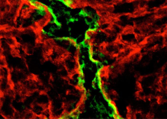
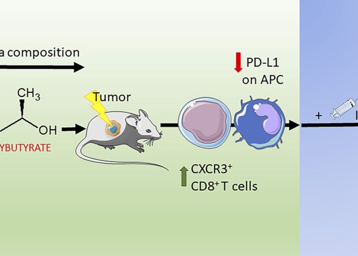
Following up with further studies, the researchers found an explanation for their observations: the new lymphatic vessels created by the melanoma tumor secrete the chemokine CCL21, which actively attracts naïve (undifferentiated) T cells into the tumor’s immunosuppressed microenvironment. Once in the tumor, these naïve T cells are locally activated following immunotherapy-induced tumor cell death, which triggers a positive feedback loop of long-lasting anti-tumor immunity.

The team tested their hypothesis on different mouse models of melanoma using multiple immunotherapy approaches, including vaccination and adoptive T cell transfer. All of these eradicated or slowed down the growth of primary melanoma tumors, and in some settings even conferred the mice with long-term protection against metastasis.

Beyond the pre-clinical models though, the scientists tested their hypothesis in human patients with melanoma. “The difference was really striking,” says Melody Swartz. “Almost all of the patients with higher than average VEGF-C levels in their blood responded to immunotherapy. This not only resulted in eradication of the primary tumors, it also encouraged T cell infiltration into metastatic tumors and resulted in long-term protection.” The researchers therefore propose that VEGF-C can be a predictive biomarker that indicates how well a patient is responding to immunotherapy.

“We now appreciate the numerous mechanisms of immunosuppression that a T cell-inflamed tumor develops to survive, including lymphangiogenesis,” the authors write. “But when the scales are tipped toward activating factors dominating over suppressive ones, as is the case with immunotherapy, these T cells become robust participants in antitumor immunity.”

The findings reveal an unappreciated role of tumor-associated lymphangiogenesis in shaping the tumor’s immune microenvironment, and pave the way for therapeutic strategies that exploit it. The study is of critical importance to cancer immunotherapy, which is growing into a formidable weapon against tumors of different types. For example, the FDA recently approved CAR-T cell therapy for leukemia and several checkpoint inhibitors are already approved for the treatment of solid tumors such as melanoma. All of these are breakthrough medicines that are now curing patients who would previously have had low chances for survival.

Story source: EPFL News

Article: Tumor lymphangiogenesis promotes T cell infiltration and potentiates immunotherapy in melanoma. Science Translational Medicine (2017). Fankhauser, M., Broggi, M. A. S., Potin, L., Bordry, N., Jeanbart, L., Lund, A. W., et al.

http://doi.org/10.1126/scitranslmed.aal4712


Continue Reading

Related Articles